Fluorescence Lifetime Imaging microscopy (FILM) is an imaging technique which is based on the differences in the exponential decay rate of the fluorophore from a sample. This technique has been widely used to quantify froster resonance energy transfer (FRET) in cells and to acquire information about populations of interacting molecules. FILM is a highly sensitive technique for recording very weak signals with picosecond resolution with high precision. It is independent of fluorophore concentration, excitation intensity and noise.
Since fluorescence lifetime depends on the molecular environment and not concentration, it can be used to map out environmental factors within cells such as PH or hydrophobicity. Combining with FRET can be used to determine protein interactions, binding interactions between large molecules, structural conformation, and various environmental effects.
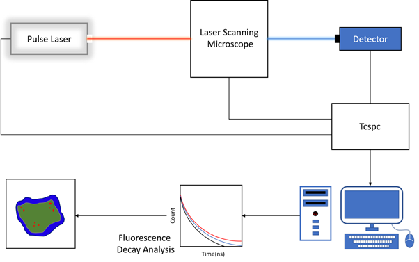
FLIM is generally based on a laser scanning microscope in combination with time-correlated single-photon counting (TCSPC), which is used to determine the fluorescence lifetime. A pulse laser system is used to illuminate the sample on a laser scanning microscope. The microscope sends the photon to a single-photon detector. A TCSPC system is connected and synchronized with all these other units. The laser system triggers the TCSPC with start signal and the detector with the stop signal. If fluorescence is detected after excitation, the TCSPC system compiles the data and send it to the computer which analyses and present the decay analysis and coloured fluorescence lifetime image. Fluorescence lifetime is determined by fitting the histogram of count versus time, by an exponential function which can consist of single- up to multi-exponential terms. When analysing the large area of the image, multi-exponential decay is often observed indicating the presence of molecule with different fluorescence lifetime. Software analyses and separates the different component of decay by fitting the intensity averaged fluorescence lifetimes for every pixel. For acquiring a fluorescence lifetime image, the emitted photons have to be attributed to the different pixels, which is performed by storing the absolute arrival times of the photons additionally to the relative arrival time in respect to the laser pulse. Line and frame marker signals from the scanner of the confocal microscope are additionally recorded to sort the time stream of photons into the different pixels. The fluorescence lifetime images are built up pixel by pixel and displayed during acquisition. The start-stop times between laser pulse and the arrival of the first photon are recorded and the average lifetime is used to reconstruct images by iterating processes for each pixel. The resultant image is displayed in a pseudocolour map.

The essential components of a FLIM set-up are: pulsed laser source, highly single photon detector, TCSPC unit to measure the fluorescence lifetime, scanning confocal microscope, dichroic mirror, objective and essential optics
SIMTRUM offers a broad variety of components that can be used in FLIM microscopy.