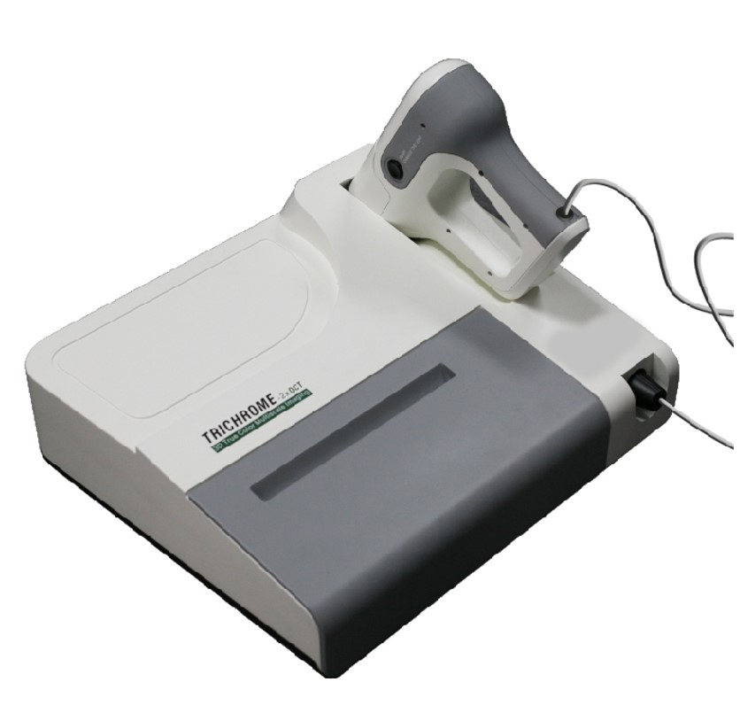
TRICHROME Series pre-clinical SD-OCT
Ultrahigh resolution and collagen fibre contrast Simtrum is proud to release TRICHROME SD-OCT which provides unprecedented spatial resolutions and tissue contrast. It is the highest resolution Optical Coherence Tomography device for preclinical use and is equipped with the unique capability of detecting collagen fiber by capturing their natural colour.
Features
- Ultrahigh axial resolution (2.5 µm in air and 1.8 µm in water); - Unique collagen fiber imaging capability based on entrinsic colour; - Handheld dermatoscopy for pre-clinical use; - Quick switch between the objective lens of various magnifications.
| 
Product Brochures : Download |
| TRICHROME - 2x OCT | Specification |
| Typical application | Research and Pre-clinical (dermatology) |
| Centre wavelength | 850 nm |
| Penetration depth | 1 mm |
| Axial resolution | 2.5 µm air and 1.8 µm in water |
| A-line rate | 68 kHz |
| Sensitivity | 105 dB @ 20 kHz scan rate A-line |
| Pixel number | 2048 or 4096 |
| Compatible scanner | Desktop scanner or handheld dematoscope |
| TRICHROME -2x OCT Objective lens selection guide |
| Focal Length | Working Distance | Spot Size | Max. Field of View | Effective Field of View |
| 4X anti-reflection coated achromat (650 - 1050 nm) |
| 50mm | 40mm | 6.3μm | 13 X 13 mm2 | 5 X 5mm2 |
| 10X NIR Long working distance Apochromat (400 - 1100 nm) |
| 20mm | 30mm | 2.5μm | 5 x 5mm2 | 2 X 2mm2 |
| 20X NIR Long working distance Apochromat (400 - 1100 nm) |
| 10mm | 20mm | 1.3μm | 2.5 X 2.5mm2 | 1 X 1mm2 |
Applications
Optical coherence tomography (OCT) is a non-invasive imaging technology, that provides real-time and cross-sectional images or fast 3D images of samples. OCT works similar to B-mode ultrasonic imaging. However, spatial resolutions of OCT can be as good as 1-2 μm, which is two orders of magnitude higher than those of ultrasound. The penetration depth of OCT is in the range of 2-3 mm. The non-contact and non-invasive nature makes OCT a perfect tool for diagnosing diseases in mucosa and surface inspection of products.
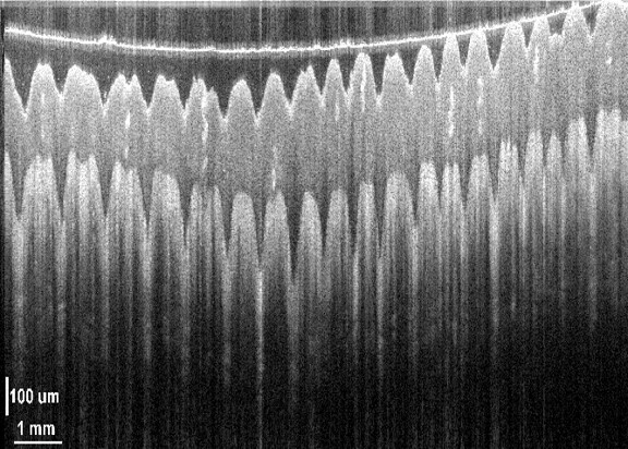
Human skin in vivo
| 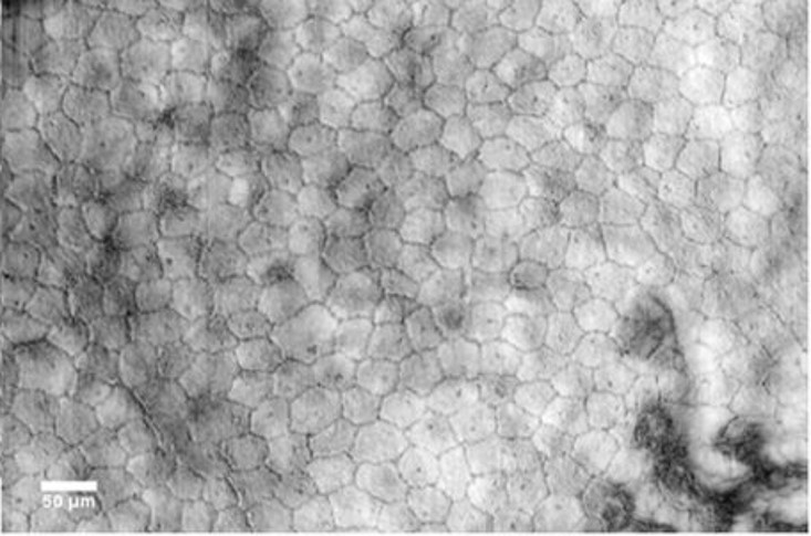
Corneal endothelium
|
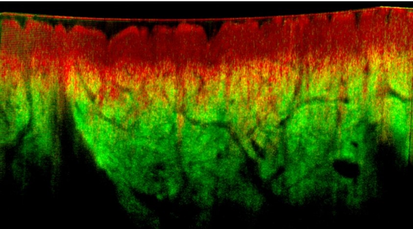
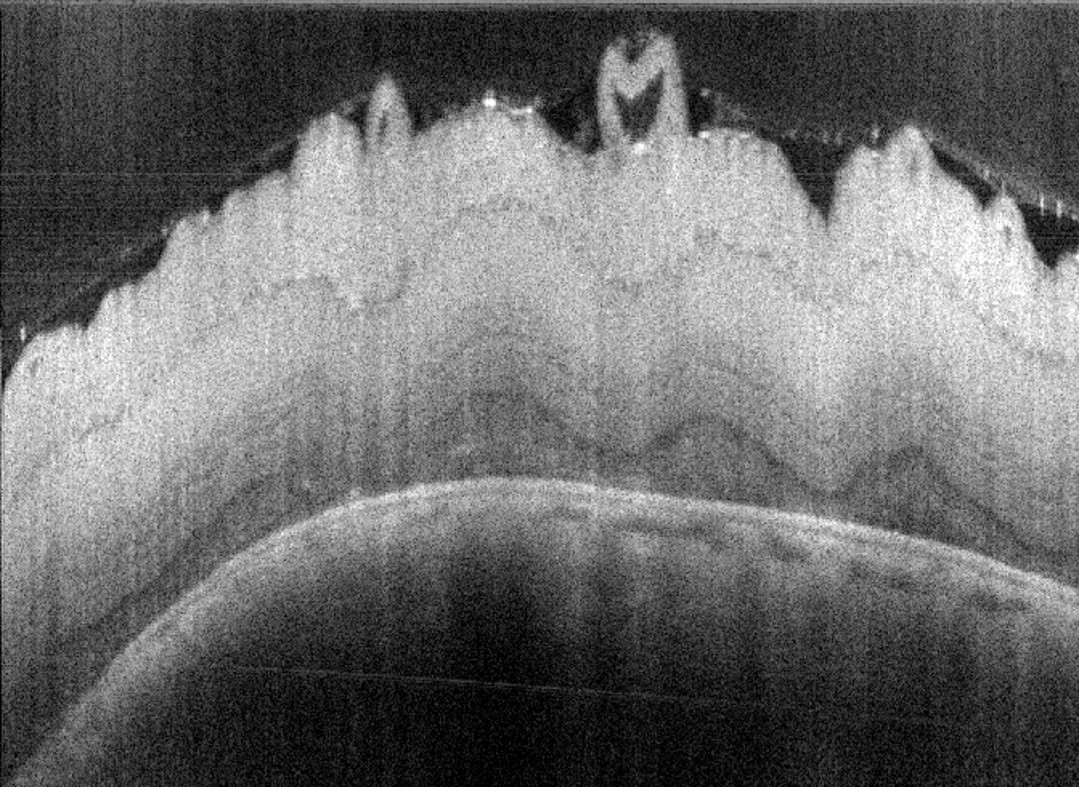
OCT colour image of the human skin in vivo

| 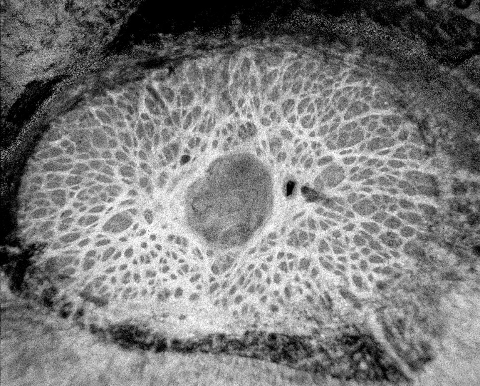
Lamina cribrosa
|
| 
Opctic nerve disc |
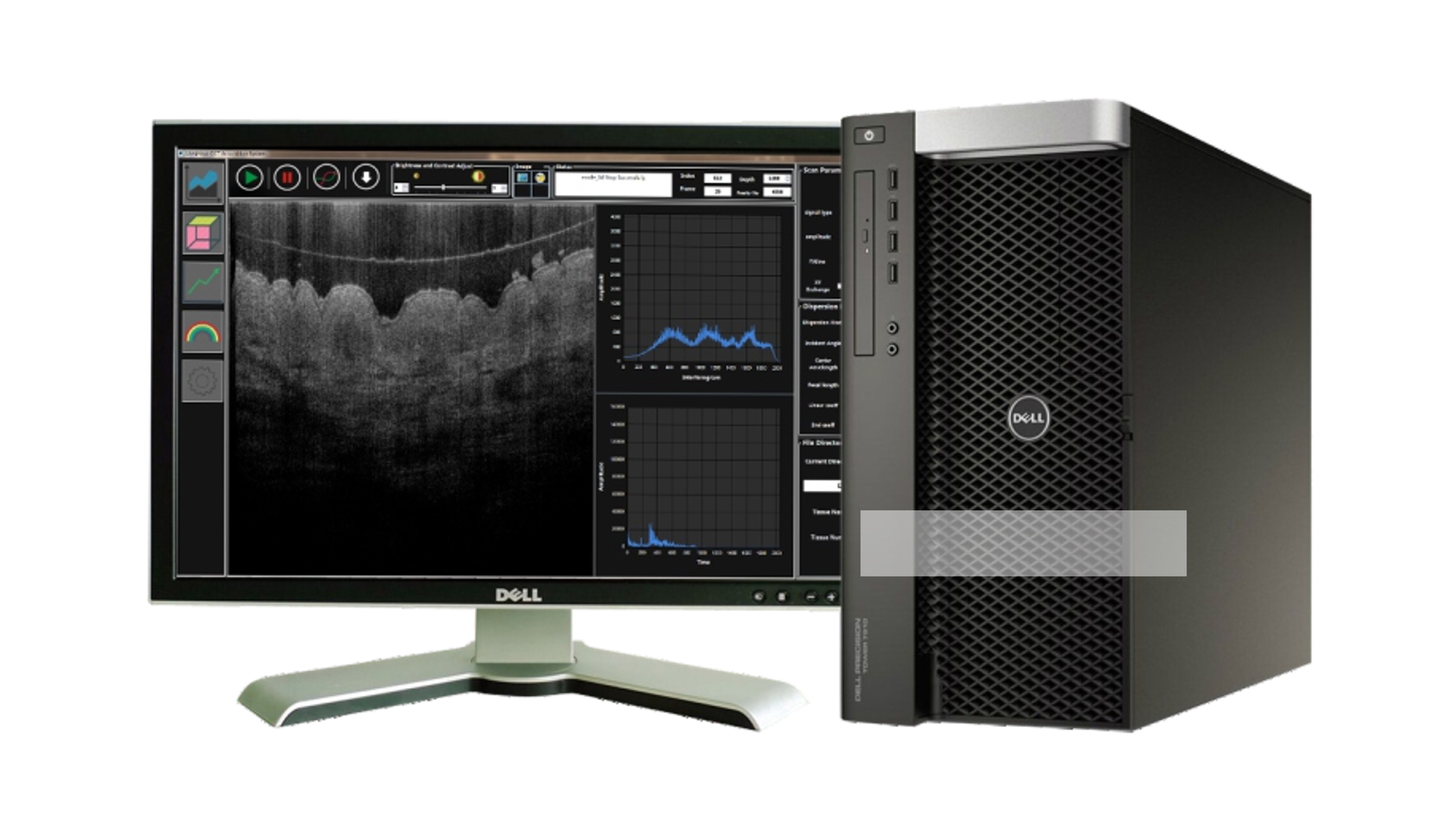
Graphics and Workstation
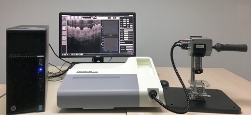
Product demonstration